
What is pericardiocentesis?
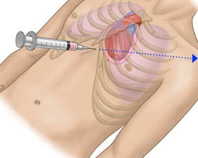
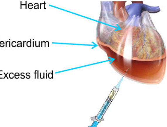
Pericardiocentesis is a procedure in which a needle is inserted through the chest wall into the pericardial space (the area surrounding the heart) to remove excess fluid. This helps relieve pressure on the heart and can improve heart function and symptoms.
Why is pericardiocentesis performed?
Pericardiocentesis is performed for several reasons:
- Relieve Symptoms: To alleviate symptoms caused by fluid accumulation, such as shortness of breath, chest pain, or swelling.
- Diagnose Underlying Conditions: To obtain fluid samples for analysis, which can help diagnose conditions causing the effusion, such as infections, cancer, or autoimmune diseases.
- Prevent Complications: To prevent or treat cardiac tamponade, a condition where fluid accumulation compresses the heart and impairs its ability to pump blood effectively.
What conditions can lead to a need for pericardiocentesis?
Conditions that may lead to pericardiocentesis include:
- Pericardial Effusion: Accumulation of fluid in the pericardial space.
- Cardiac Tamponade: Severe compression of the heart due to fluid accumulation.
- Infections: Such as bacterial or viral pericarditis.
- Cancer: Tumors or malignancies affecting the pericardium.
- Autoimmune Diseases: Conditions like systemic lupus erythematosus or rheumatoid arthritis.
- Trauma: Injury to the chest or heart.
How is pericardiocentesis performed?
The procedure typically involves the following steps:
- Preparation: The patient is positioned, usually sitting up or lying on their back. Local anesthesia is administered to numb the area where the needle will be inserted.
- Imaging Guidance: The procedure is often guided by ultrasound or fluoroscopy (X-ray) to locate the fluid and ensure accurate needle placement.
- Needle Insertion: A needle is inserted through the chest wall, typically below the sternum or on the left side, into the pericardial space.
- Fluid Aspiration: Fluid is withdrawn through the needle into a collection bag or syringe.
- Post-Procedure Care: The needle is removed, and pressure is applied to the insertion site. The patient is monitored for any complications.
